Sight is one of our most essential senses, and protecting it should remain a top priority. One serious condition that can threaten your eyesight is retinal detachment—a medical emergency that requires immediate attention. If not addressed promptly, it may result in irreversible loss of vision. In this article, we’ll explore what retinal detachment is, the causes, warning signs, treatment options, and how you can protect your healthy vision.
What Is Retinal Detachment?

Retinal detachment happens when the retina—a delicate layer of tissue at the back of the eye that detects light and sends visual signals to the brain—separates from its usual position. When this happens, the retina can no longer function properly, which puts vision at risk.
Retinal detachment is classified into three primary types:
- Rhegmatogenous – The most common type, caused by a tear or break in the retina.
- Tractional – Occurs when scar tissue pulls on the retina, often linked to diabetes.
- Exudative – Results from fluid accumulating beneath the retina without any tear, typically triggered by inflammation or eye injury.
What Causes Retinal Detachment?
Multiple factors can raise your chances of experiencing a detached retina:
- Aging – The retina naturally becomes thinner and more fragile over time
- Severe myopia (nearsightedness) – Increases stress on the retina
- Eye injury or trauma
- Previous eye surgery (like cataract removal)
- Family history of retinal detachment
- Diabetic eye disease
If you’re in a high-risk group, regular eye exams are essential for maintaining healthy vision and catching issues early.
Warning Signs and Symptoms
Retinal detachment is often painless, which makes it even more important to know the warning signs. Symptoms can develop abruptly and may involve:
- A sudden increase in floaters (tiny specks or strings drifting in your vision)
- Bursts of light, particularly noticeable in your side (peripheral) vision
- A shadow or dark curtain drifting across your vision
- Blurred or distorted vision
- Gradual loss of peripheral (side) vision
If you notice any of these symptoms, seek emergency eye care immediately. Early diagnosis greatly improves your chances of recovery and vision preservation.
How Is Retinal Detachment Diagnosed?
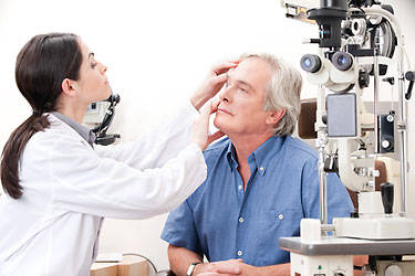
An eye care professional (optometrist or ophthalmologist) can diagnose retinal detachment through a comprehensive eye exam. Common tests include:
- Dilated eye examination – Allows a clear view of the retina and the back of the eye
- Ocular ultrasound – Especially helpful if the retina can’t be seen clearly due to bleeding
- Optical coherence tomography (OCT) – For high-resolution imaging of retinal layers
Prompt diagnosis is crucial because once the retina loses connection with the back of the eye, its cells start to die quickly.
Treatment Options
The treatment approach varies based on the extent and type of retinal detachment. Common procedures include:
- Laser Surgery (Photocoagulation)
Used for small tears or holes before full detachment occurs.
- Cryopexy
Applies freezing therapy around the retinal tear to form scar tissue that helps seal and secure it in place.
- Pneumatic Retinopexy
A gas bubble is introduced into the eye to gently press the retina back into its proper position.
- Scleral Buckling
A small band is placed around the eye to gently push the wall against the detached retina.
- Vitrectomy
Removes the vitreous gel inside the eye and replaces it with gas or silicone oil to hold the retina in place.
Most of these procedures are outpatient surgeries with a reasonable recovery time, although follow-up care is essential.
Recovery and Aftercare
Recovery can vary depending on the procedure but may include:
- Wearing an eye patch or shield
- Limiting physical activity and avoiding lifting
- Maintaining a specific head position (especially after gas bubble treatments)
- Regular follow-up appointments to monitor healing
Avoiding smoking, managing chronic conditions like diabetes, and staying on top of medications will also support the healing process and future eye health.
Preventing Retinal Detachment
Although not every case can be avoided, you can reduce your risk by taking the following precautions:
Schedule regular comprehensive eye exams, particularly if you’re over 50 or have severe nearsightedness
Use proper eye protection during sports or activities with a high risk of injury
Keep chronic conditions like diabetes and high blood pressure under control.
Watch for and respond quickly to any sudden vision changes.
Follow up after eye surgeries or trauma, even if you feel fine
Final Thoughts
Retinal detachment is a serious eye condition that demands quick action. By knowing the signs and maintaining regular check-ups, you can greatly reduce your risk of permanent vision loss.
Protecting your healthy vision starts with awareness and proactive care. Whether you’re noticing new symptoms or simply want to stay ahead of potential problems, early intervention is key to long-term eye health.


